Figure 1.
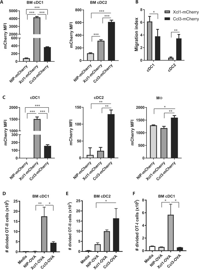
Characterization of Ccl3- and Xcl1-fusion vaccines. (A) BM DCs were incubated for 18 h with 0.5 μg Ccl3-, Xcl1- or anti-NIP-mCherry, and specific staining of cDC1 and cDC2 evaluated by flow cytometry after gating as indicated in Supplementary Fig. S1B. (B) Chemotaxis of BM DC were evaluated in transwell plates with 1.5 μg/ml Ccl3-, Xcl1- or anti-NIP-mCherry added to the bottom well. Migrated cells were identified as cDC1 or cDC2 by flow cytometry. The number of migrated cells were normalized to the number of spontaneously migrated cell when only medium was added to the bottom well. (C) In vivo targeting of APC. BALB/c mice were injected i.v. with 20 μg Ccl3-, Xcl1- or anti-NIP-mCherry. Spleens were harvested after 2 h and the mCherry staining of cDC1, cDC2 and macrophages analyzed by flow cytometry after gating as described in Supplementary Fig. S1D. (D,E) Proliferation of OT-II (D) and OT-I (E) cells after 4 days incubation with cDC1 or cDC2 and NIP-, Ccl3- or Xcl1-OVA. Number of proliferating cells was determined by CTV dye dilution by flow cytometry. Data shown are mean + SEM and representative of 2 independent experiments with (A) 6 replications or (B,D,E) 3 replications pr. group, or (C) 3 mice pr. group. Statistical analysis performed using (A,C) one-way ANOVA with Tukey’s multiple comparison test, (B) t-test, *p < 0.05, **p < 0.01, ***p < 0.001.

