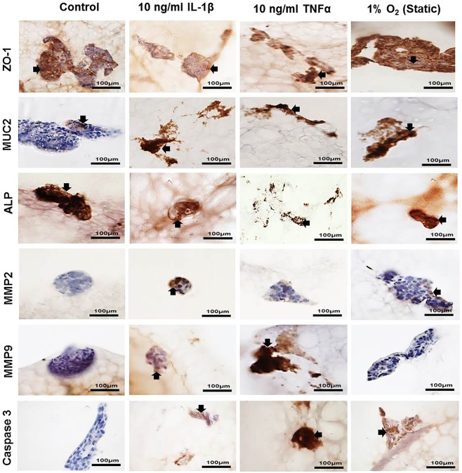
Figure 7.
Immunopositivity (brown) of co-culture Caco-2 and HT29-MTX cells at percentages 90% Caco-2/10% HT29-MTX layered on L-pNIPAM hydrogel scaffolds under dynamic culture conditions following 12 weeks as control or for 11 weeks and then treated with 10 ng/ml IL-1β or 10 ng/ml TNFα for 1 week under dynamic culture conditions or hypoxic at 1% O2 for 1 week under static culture conditions. Cell nuclei were stained with haematoxylin (blue). The black arrows indicate positively stained cells. Scale bar = 100 µm

