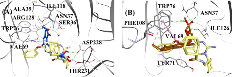
Fig. 3. Molecular docking models for the mixed-type and the noncompetitive BACE1 inhibitors.
Molecular docking models for a the mixed-type BACE1 inhibitor (2R,3R)-pteroside C (blue color) and b the noncompetitive BACE1 inhibitor (2R)-pteroside B (brown color). Docked poses are superimposed on the X-ray crystal structure of QUD (yellow color) (PDB code: 2WJO). BACE1, active site residues and compounds are shown by ribbon, line and stick models, respectively. Colors of the dotted lines explain the types of various interactions: hydrogen bonding interactions (green), hydrophobic interactions (pink) and π-sigma interactions (purple). BACE1 β-site amyloid precursor protein cleaving enzyme 1

