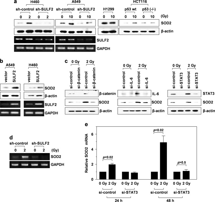
Fig. 1. IR induces SOD2 expression via the p53/SULF2/β-catenin/IL-6/STAT3 pathway.
a–d Western blotting and RT-PCR were performed 48 h after γ-irradiation. a H460 and A549 lung cancer cells (p53wt) were infected with lentiviruses expressing control (nontargeting sequence) or SULF2-specific shRNA. These transfectants, along with H1299 lung cancer cells (p53null) and p53wt-expressing or p53-knockout HCT116 colon cancer cells, were irradiated with the indicated doses of γ-rays, and SOD2 levels were compared by western blot analysis using β-actin as a loading control. SULF2 expression was compared by RT-PCR using GAPDH as a loading control. b A549 and H460 cells were transfected with an empty or SULF2 expression vector, and SOD2 protein and SULF2 mRNA levels were compared. c H460 cells treated with a control or an siRNA targeting β-catenin, IL-6, or STAT3 were irradiated with 2 Gy of γ-rays, and the levels of the indicated proteins were compared. d H460 cells infected with the lentiviruses indicated in a were irradiated, and SOD2 mRNA levels were analyzed by RT-PCR. e H460 cells treated with a control or a STAT3-targeting siRNA were irradiated, and SOD2 mRNA levels were compared by quantitative real-time PCR at 24 and 48 h after irradiation

