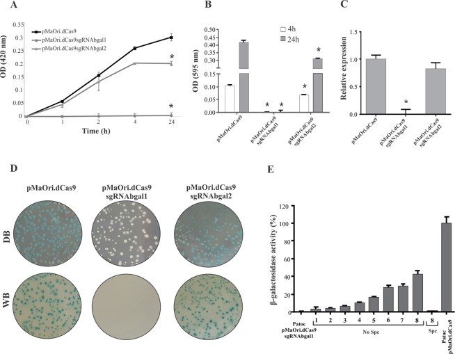
Figure 5.
Total β-galactosidase gene silencing obtained by dCas9 expression along with sgRNA pairing to the coding strand. Different colonies were selected from the EMJH plates, grown in liquid media and then evaluated regarding β-galactosidase activity in a time-basis, employing ONPG substrate (A) and X-gal (B). Graphics show the average of densitometric reading plus standard deviation of 4 biological replicates. For statistical analysis, absorbance values displayed by cells containing dCas9 and sgRNA were compared to those displayed by cells containing pMaOri.dCas9 plasmid alone by Student’s two-tailed t test (*P < 0.05). (C) Relative transcription level of β-galactosidase gene in knockdown strains by qPCR. Presented values represent the mean plus standard deviation measurements. For statistical analyses, the absorbance or relative expression values displayed by the knockdown mutants were compared to those presented by the cells containing pMaOri.dCas9 alone (*P < 0.05). (D) X-gal solution was spread onto EMJH plates containing grown colonies, containing either pMaOridCas9 or this plasmid with sgRNA cassettes. Figures are presented against dark (DB) and white (WB) background. (E) L. biflexa cells containing pMaOri.dCas9sgRNAbgal1 grown in EMJH plus spectinomycin were transferred to new EMJH liquid medium (1:10 dilution) with no antibiotic (No Spc). Serial passages were performed (1–8) and at each passage, β-galactosidase activity was measured. The activity displayed by cells containing pMaOri.dCas9 plasmid was considered 100%.

