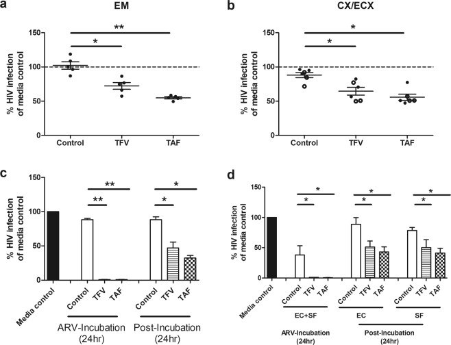
Figure 4.
Effect of fibroblasts and endometrial (EM) epithelial cells in decreased HIV infection of CD4+ T cells. Conditioned media (CM) was collected from (a) endometrium (EM), (b) CX/ECX fibroblasts pre-treated with TFV (3277 µM) or TAF (10 µM) for 24 hr. CM were collected 24 hr post ARV washout and incubation with fresh media; CM was incubated with activated CD4+ T cells prior to HIV infection. Secreted p24 levels in the culture media after 5 days of infection were measured by p24 ELISA as described in Methods. CM was collected from EM fibroblasts of 5 patients while CM from CX (dark circle) and ECX (open circle) fibroblasts was from 3 matched patients. Each circle represents an individual patient. Data are normalized in (a,b) to the infection of CD4+ T cells in the absence of CM (media control) and set to 100%. Each circle represents a different patient. Blood CD4+ T cells were isolated from 4 donors. Horizontal lines represent the mean and SEM respectively. *p < 0.05. **p < 0.01. (c) Polarized EM epithelial cells were apically treated with TFV or TAF for 24 hr, after which basolateral CM was immediately collected (ARV-Incubation). Following rinsing, cells were incubated with fresh media (no TFV or TAF) for an additional 24 hr prior to CM collection (Post-Incubation 24 hr). Activated CD4+ T cells from blood were incubated with CM for 24 hr and washed and infected with HIV for 2 hr. Secreted viral p24 levels were measured after 5 days of infection (Methods). Data are normalized to the infection of CD4+ T cells in the absence of CM (media control) which is set to 100%. Bars represent EM tissues from 3 patients. (d) Effect of epithelial cells and fibroblasts on prevention of HIV infection. Polarized EM epithelial cells grown in the upper chamber of cell inserts were treated apically with TFV (3277 µM) or TAF (10 µM) for 24 hr in the presence of confluent fibroblasts (SF) from the same donor grown in the lower chamber (no cell contact). Following incubation, epithelial cells and stromal fibroblasts (EC + SF), basolateral CM was collected for analysis (ARV-Incubation). Following extensive washing to remove extracellular ARVs (Post-Incubation), epithelial cells alone (EC) and stromal fibroblasts alone (SF) were incubated in fresh media for 24 hr, after which CM were collected. CD4+ T cell protection against HIV infection for each CM was measured as described in Methods. Data are normalized to the infection of CD4+ T cells in the absence of CM (media control). Bars represent 4 patients. Blood CD4+ T cells were isolated from 2 donors. Column and horizontal lines represents the mean and SEM respectively. *p < 0.05. **p < 0.01.

