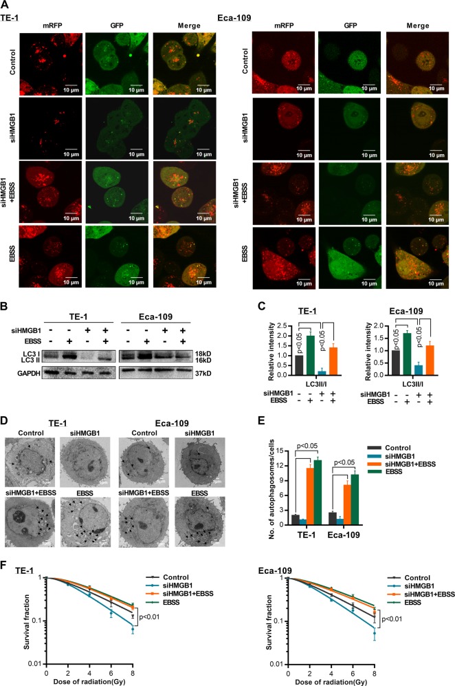
Fig. 5. Activation of autophagy reverses radiosensitization induced by HMGB1-knockdown.
a ESCC cells stably expressed mRFP-GFP-LC3 protein were transfected with siHMGB1 or treated with starvation (EBSS for 4 h). Laser confocal fluorescence microscopy analyses for autophagosomes and autophagosomes. In the merged image, yellow puncta indicate autophagosomes, while red puncta indicate autolysosomes. b Western blot was performed to detect LC3 I, LC3 II, and HMGB1 protein expression in siHMGB1-transfected TE-1 or Eca-109 cells and treated with starvation (EBSS for 4 h). c Relative protein density according to GAPDH was analyzed by NIH-Image J, t test. d TEM was used to observe the autophagosomes in siHMGB1-transfected TE-1 or Eca-109 cells and treated with starvation (EBSS for 4 h). e The number of autophagosomes in TE-1 and ECA-109 cells under different conditions. f Survival curves of TE-1 and Eca-109 treated with IR (0, 2,4,6,8 Gy)

