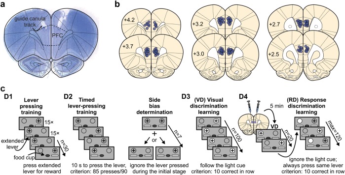
Fig. 1.
Histology and set-shift procedure. a Microphotograph of Cresyl violet stained coronal section containing guide canulae tracks for injections directly in the prefrontal cortex (PFC). b Schematic representation of the injection sites (circles) superimposed on the coronal plane of a rat brain (modified from [25]). Numbers on the schemes indicate distance from Bregma point. c Experimental timeline for the attentional set-shift procedure. The grey rectangles represent front panel of the operant box, circles correspond to the light cues (gray = light off; white = light on), white rectangles correspond to extended levers, the lever rewarded with sucrose pellet is indicated with plus sign above it; D1-D4 indicate different days of the procedure. Note that rats were injected with the drug solution on the last day of the procedure

