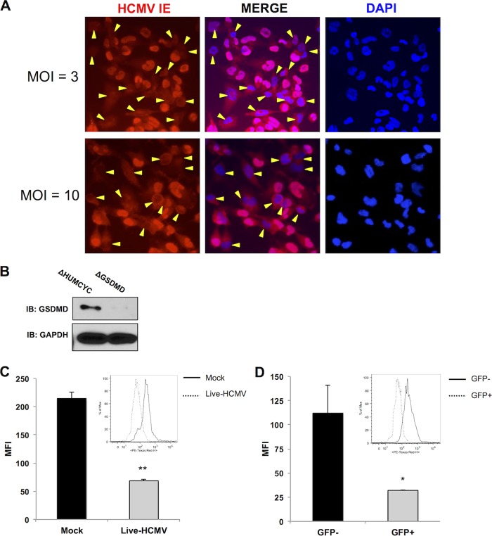
FIG 8.
Levels of pro-IL-1β are reduced in HCMV-positive cells. (A) Immunofluorescence microscopy showing HCMV IE expression in THP-1 cells infected at the indicated MOI overnight. Yellow markers indicate DAPI-stained cells that lack detectable nuclear IE protein. (B) Immunoblot assay for GSDMD in ΔHUMCYC and ΔGSDMD THP-1 cells. (C, D) Intracellular staining for IL-1β in ΔGSDMD EF1α-pro-IL-1β THP-1 cells either mock infected or infected with HCMV-TB40/E (MOI of 3) for 18 h (C) and gated for virally encoded GFP (D). GFP-positive (GFP+) population depicts the number of HCMV-infected cells in the sample. The bar graphs show the mean fluorescence intensities (MFI) for IL-1β staining. The insets show IL-1β staining in the indicated populations. Values are averages ± SEM from two biological replicates. Student’s t test was performed. *, P < 0.05; **, P < 0.01.

