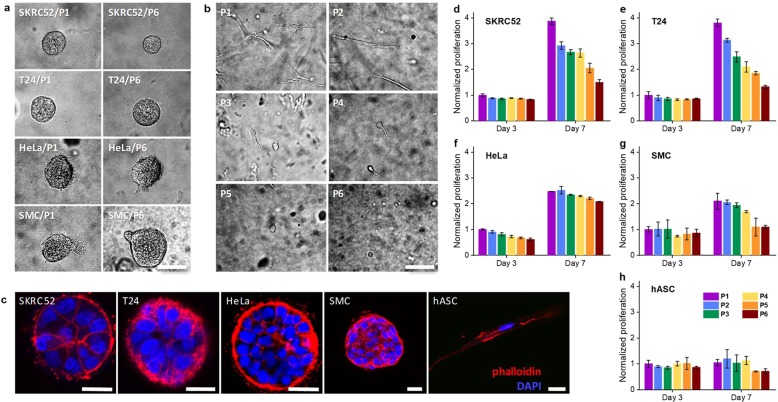
Figure 3.
Cell behavior in PIC gels with different GRGDS densities based on P1–P6. (a) SKRC52s, T24s, HeLas, and SMCs in P1 and P6 after 7 days of culturing. Irrespective of the peptide density, cell spheroids are formed for all cell types. (b) hACSs show strongly different morphologies in P1–P6 after 7 days of culturing: elongated morphologies at the highest GRGDS densities and spherical morphologies at low densities. (c) Fluorescence staining with Phalloidin (red, F-actin) and DAPI (blue, cell nuclei) of different cell types after 7 days of culturing in P2 show that the multicellular aggregate differs in nuclear and cytoskeletal arrangement. Contrarily, hACSs show single elongated morphologies. (d–h) Proliferation, normalized to absorbance of P1 at day 3, for different cell types after 3 and 7 days of culturing in P1–P6. SKRC52s, T24s, HeLas, and SMCs show increased proliferation with higher peptide densities. For hASCs, proliferation in all gels is low. Note that the colors in d–h match those in Figure 2a. For all samples, the PIC concentration c = 2 mg mL–1 and cell density is 20000 cells mL–1. The scale bar for all figures is 70 μm. The error bars in d–h represent standard deviations of three experiments.

