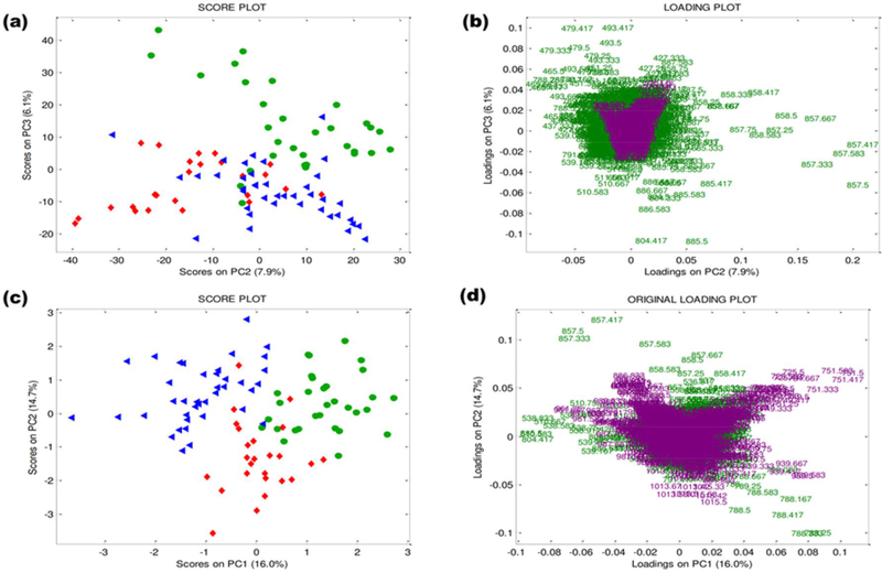
Fig. 3. PCA of the fused datasets from positive and negative ion mode mass spectra in a low-level approach:
(a) PC2 vs. PC3 score plot. (b) PC2 vs. PC3 original loading plots labeled in terms of m/z ratio. PCA of the fused datasets from positive and negative ion mode mass spectra in a mid-level approach: (c) PC1 vs. PC2 score plot. (d) PC1 vs. PC2 original loading plots labeled in terms of m/z ratio. Immature oocytes: green circles (n = 31), 24-hour in vitro matured oocytes: red diamonds (n = 25), 44-hour in vitro matured oocytes: blue triangles (n = 40). Negative ions: green; positive ions: violet.

