Fig. 12.
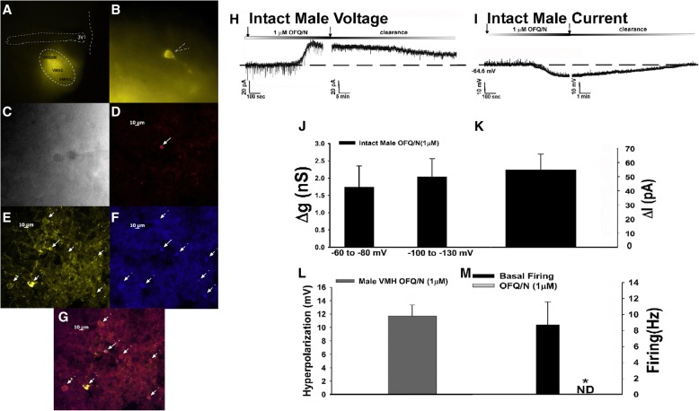
The N/OFQ-induced activation of GIRK channels occurs in SF-1 neurons located in the VMN of NR5A1-Cre mice. a YFP labeling of SF-1 neurons at × 4 magnification. b YFP labeling of the recorded SF-1 neuron at × 40 magnification. c DIC image of the previous neuron taken during recording. d Biocytin labeling of the cell in (c) visualized with streptavidin/AF546 as indicate by solid line. e YFP labeling of the same cell seen in (b–d). Surrounding YFP-filled neurons are indicated by dashed lines. f An antibody directed against SF-1 immunolabels the cell in c as visualized with AF350. g A composite overlay of the biocytin/YFP/SF-1 labeling seen in the cell. Panels d–g were photographed at × 20. The calibration bar equals 10 μm. Membrane current traces showing the N/OFQ-induced outward current (h; n = 5) and hyperpolarization (i; n = 8) in SF-1 neurons. The robust outward current is accompanied by an increase in slope conductance (j and k), and the hyperpolarization is associated with a decrease in firing (l and m). *p < 0.05, Student’s t test

