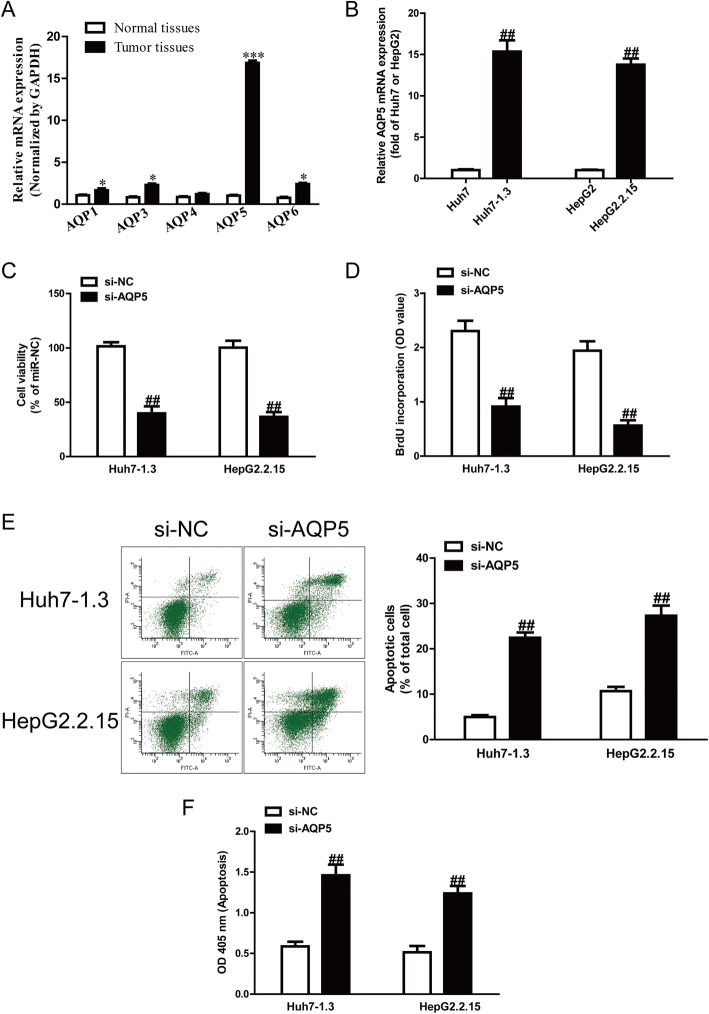
Fig. 1.
Expression of AQP5 and its effects on cell proliferation and apoptosis of HBV-HCC cells. a mRNA and protein expression of AQP1, AQP3, AQP4, AQP5 and AQP6 in normal liver tissues (n = 20) and HBV-HCC tissues (n = 20) was detected by qRT-PCR. b mRNA expression of AQP5 in HepG2, HepG2.2.15, Huh7 and Huh7–1.3 cells. Cell proliferation was assessed by CCK-8 assay (c) and BrdU-ELISA assay (d). Cell apoptosis was measured by flow cytometric analysis of cells labeled with Annexin-V/PI double staining (e) and nucleosomal degradation using Roche’s cell death ELISA detection kit (f). The data shown are mean ± SEM, n = 4. *P < 0.05, ***p < 0.001 vs. normal tissues; ##p < 0.01 vs. HepG2, Huh7 or si-NC

