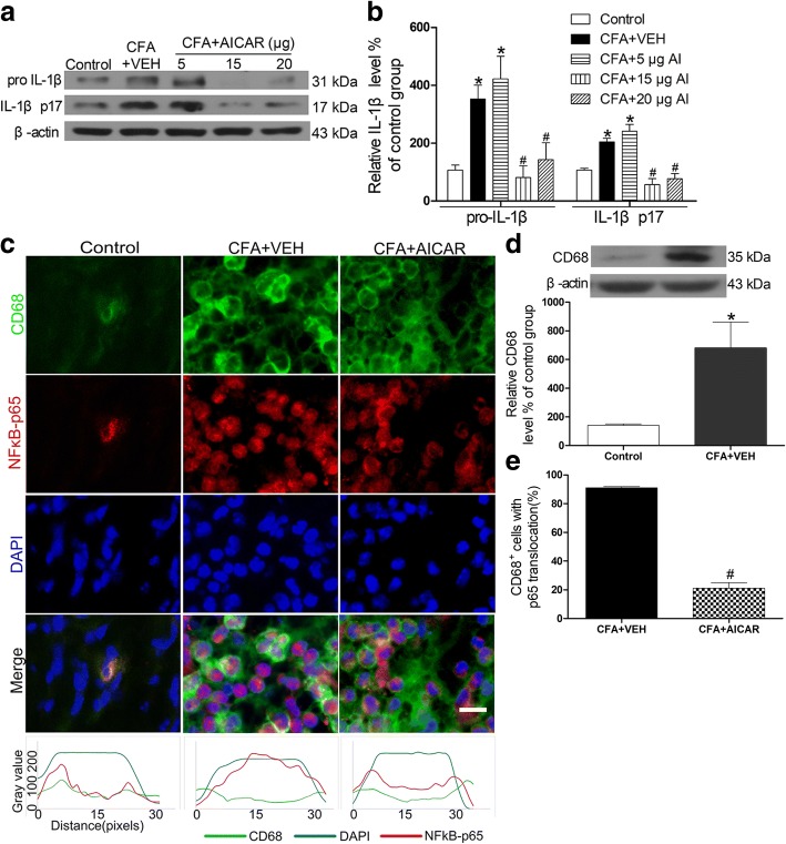
Fig. 2.
AICAR downregulates expression of IL-1β and suppresses CFA-induced NF-κB activation in macrophages of the inflamed skin. AICAR (5, 15, 20 μg/20 μl) was administrated at CFA injection day 4. Skin tissues were collected at 2 h after AICAR treatment for Western blotting and immunofluorescence. Representative gels and quantification for pro-IL-1β and IL-1β p17 bands (a and b) are shown. One-way ANOVA revealed a significant difference at *P < 0.05 vs. Control group and #P < 0.01 vs. CFA+VEH group. c AICAR inhibited the CFA-induced NF-κB translocation from the cytosol to the nucleus, as demonstrated by the immunofluorescence of p65 in CD68 marked macrophage cells. Scale bar represents 50 μm. The bottom line of the panels shows the merged profiles of the fluorescent intensity of CD68, NF-κB p65 and DAPI signals along the lines drawn through the axis of immunostaining macrophage cells. d Quantification for CD68 protein level of skin tissues. e Quantification for cells with p65 translocation of c. Non-paired Student’s t test revealed a significant difference at *P < 0.05 vs. Control group and #P < 0.01 vs. CFA + VEH group (n = 4 mice/group). The abbreviations used here are VEH (vehicle of AICAR) and AI (AICAR)

