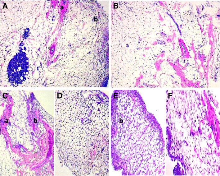
Fig. 7.
Microphotograph of H&E-stained histological section of the synovial membrane of rats (40x magnification). a zymosan control: intense leukocyte infiltrate with fibrosis area. b normal control: structures preserved and within normality. c LfAE 100 mg/kg with presence of leukocyte infiltrate and areas of fibrosis. d LfAE 200 mg/kg presenting reduction of inflammatory parameters in the synovial membrane. e LfAE 300 mg/kg group presenting mild leukocyte infiltrate in the synovium. f Standard 100 mg/kg with mild leukocyte infiltrate. a - fibrosis; b - inflammatory infiltrate; c – neovascularization

