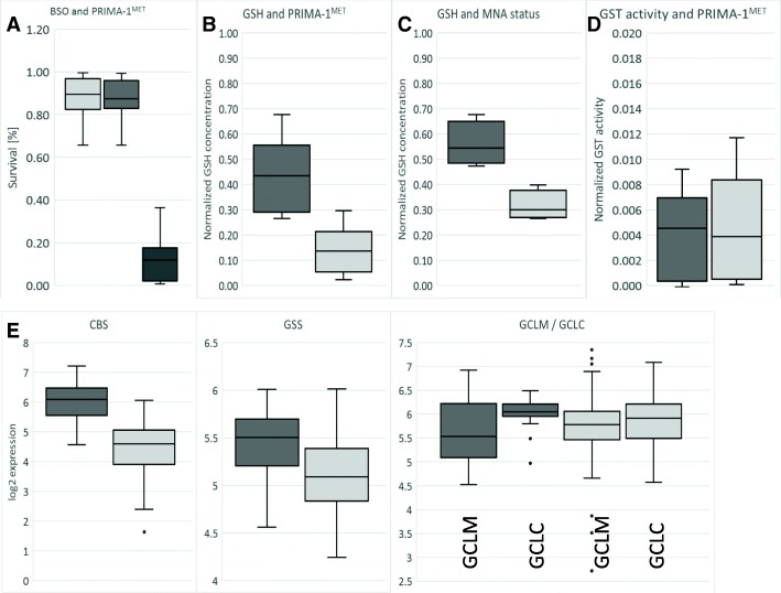
Fig. 5.
PRIMA-1MET and glutathione. a: Concurrent BSO and PRIMA-1MET treatment. All cell lines were treated with fixed 100 μM BSO for 6 h prior to PRIMA-1MET treatment at a concentration of less than IC90 on average. The Y-axis indicates the survival ratio. A significant synergistic effect (CDI = 0.16) due to BSO treatment was detected in all cell lines, indicating that the depletion of GSH significantly influences the sensitivity of the cells to PRIMA-1MET. Light grey: survival at 100 μM BSO, dark grey: survival at IC90 concentration of PRIMA-1MET, black: BSO and PRIMA-1MET. b: Concentration of GSH decreases 3.1-fold (p < 0.001) in NB cell lines treated with 60 μM PRIMA-1MET for 6 h (light grey) in comparison to non-treated (dark grey). c: Concentration of GSH is 1.8-fold (p < 0.05) lower in MNA NB (dark grey) in comparison to non-MNA NB (light grey). d: No significant difference in GST activity due to PRIMA-1MET (light grey) in comparison to non-treated control (dark grey) NB cell lines. e: Higher expression of CBS (2.97-fold, p < 0.001) and GSS (1.25-fold, p < 0.001), and significant change in GCLC/GCLM ratio (1.3-fold, p = 0.037) in MNA NB samples (dark grey) in comparison to non-MNA NB samples (light grey). Dots: outliers

