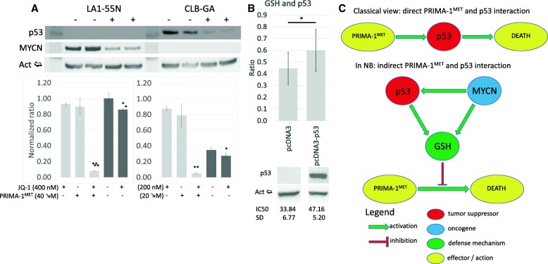
Fig. 6.
PRIMA-1MET, p53 and MYCN. a: Inhibition of MYCN and p53 expression by JQ-1. LA1–55 N (p53-null) cells showed a significant decrease in MYCN, whereas CLB-GA (non-MNA) cells demonstrated almost complete suppression of p53 expression after 16 h treatment. - – Non-treatment control, + − 16 h, 200 nM JQ-1. PRIMA-1MET and JQ-1 demonstrated synergism in both cell lines (CDILA1–55N = 0.1, CDICLB-GA = 0.07). Treatment with JQ-1 resulted in a significant decrease in the concentration of GSH in LA1–55 N (1.163-fold, p < 0.01) and CLB-GA (1.26-fold, p = 0.028). Light grey – JQ-1/PRIMA-1MET co-treatment, dark grey – GSH concentration after JQ-1 treatment, *p < 0.05, **p < 0.01. b: Concentrations of GSH and IC50 in LA1–55 cells with and without p53 gene expression. Results demonstrated 1.34-fold (p = 0.029) increase in total cellular GSH and 1.39-fold (p = 0.007) IC50 increase in cells expressing p53. Upper – concentration of GSH normalized to the number of cells using CellTiter2.0. Middle – WB for p53. Bottom – IC50 values for cells with and without p53. * - p < 0.05. c: New proposed model of interaction between PRIMA-1MET, p53, and MYCN

