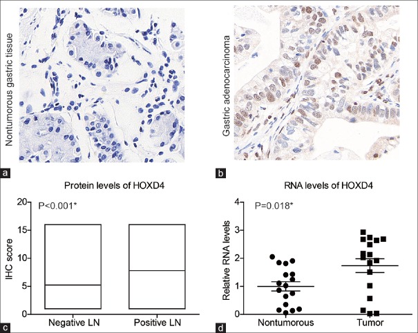
Figure 1.
Analyses of homeobox D4 expression in patients with gastric adenocarcinoma. Immunohistochemistry staining of homeobox D4 in non-tumorous gastric tissues (a) and gastric adenocarcinoma tissues (b). (c) Correlation between homeobox D4 immunohistochemistry results and tumor metastasis, showing that patients with negative lymph node had a lower homeobox D4 protein expression level compared to those with positive lymph nodes. (d) RNA levels of homeobox D4 were examined in tumor tissues and adjacent non-tumorous tissues. *P < 0.05 by Student's t-test

