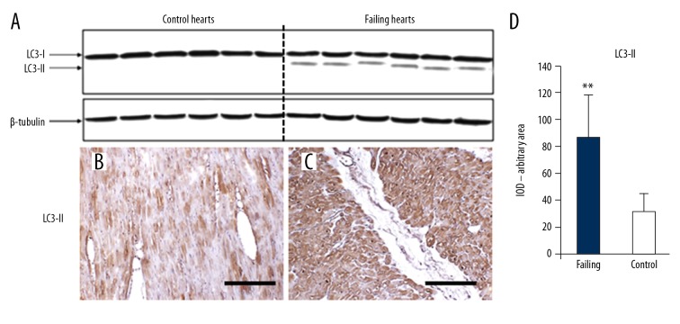
Figure 2.
(A) Western blotting analysis for LC3-I and LC3-II. Cleaved LC3 is present in failing hearts only. (B) Anti-LC3-II IHC in control hearts: the cardiomyocytes show faint staining in small cytoplasm areas. (C) In failing hearts, the cardiomyocytes appear more intensely stained; scale bar 100 μm. The optical density for LC3-II staining is resumed in (D); ** t-test P<0.000. LC3 – light-chain-3 protein; IHC – immunohistochemistry.

