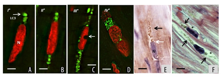
Figure 3.
Representative progression of autophagy in failing heart. (A) Stage I: LC3-II staining (green) began at one cytoplasmic nuclear pole of the cardiomyocyte; N – nucleus, propidium iodide staining. (B) Stage II: the autophagy extended to both nuclear poles. The nuclei have normal appearance. (C) Stage III: the autophagy radiated to more peripheral cytosolic areas. The nuclei exhibit findings of focal compromise in form of single or multiple concavities (arrow) as “bite-like” signs. (D) Stage IV: in the later stages, the nucleus underwent progressive vacuolization and final disintegration. Cardiomyocytes with massive green staining in combination with the red staining of nucleus, assume a “strawberry-like” appearance. (E) Representative IHC for anti-LC3-II ABC-diaminobenzidine staining shows a massive presence of stained vacuoles (black arrow) that originated from a nuclear pole (white arrow), N – nucleus. (F) Representative H and E staining of later stages (III and IV) of autophagy progression. Unipolar and bipolar clustered granules of different size (arrows), the nuclei appear vacuolated and shows small bit signs. The cytoplasm is apparently empty (asterisks); white bar=5 μm; black bar=10 μm. LC3 – light-chain-3 protein; IHC – immunohistochemistry.

