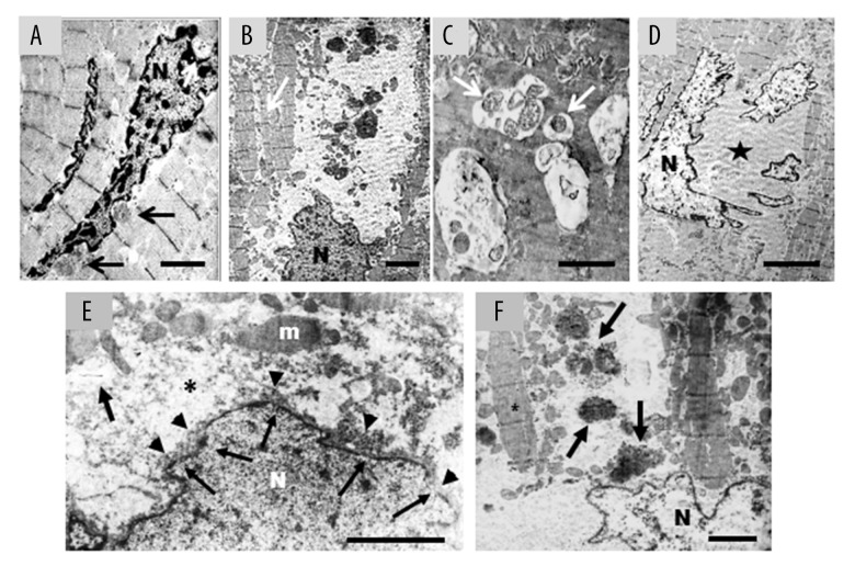
Figure 4.
(A–F) Failing heart: representative electron microscopy pictures of cardiomyocytes alteration in advanced stages of damage. (A) Damaged cardiomyocytes sometime show nuclei with chromatin condensation, combined with invaginations of the nuclear envelope (arrows) that contain finely granular material cytoplasm without organelles. It is interesting to note the close similarity between this ultrastructural image and the nuclear concavities in Figure 3C; scale bar=2.5 μm. (B) The autophagy extends from the perinuclear areas to farther portion of cytosol. This is devoid of organelles and contains few small mitochondria and voluminous electron-dense material (probably lipofuscin and/or residue of autophagosomes) of variable size immersed in finely granular and faint electron-dense matrix. The myofibrils are isolated (arrow) and shown sign of disorganization; scale bar=3 μm. (C) Dilated vesicles of variable size containing mitochondria (arrows), and/or other unrecognizable residues were commonly observed; scale bar 1 μm. (D) At later stages the nucleus is fragmented, chromatin is condensed and marginated. Large cytoplasmic area devoid of organelles is evident (star); scale bar=5 μm. (E) The nuclear envelope is irregular with small interruptions (arrows) from which light-electron-dense nuclear material comes out (head arrows). Around the nucleus, there is evident a large area of cytoplasm devoid to organelles (asterisk); m – mitochondria; scale bar=1.5 μm. (F) At very advanced stage, nuclear content appears more electron-transparent. The cytoplasm is empty and very disorganized, many waste products are present (arrows) and myofibrils are isolate (asterisk); scale bar=2.5 μm; N – nucleus.

