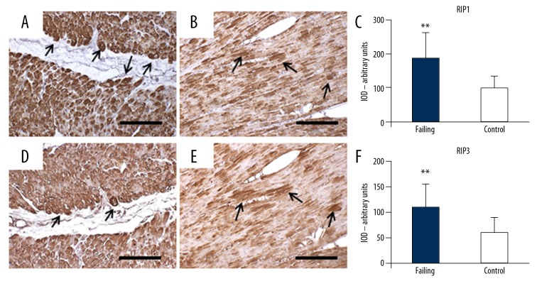
Figure 7.
Serial sections with anti-RIP1 and anti-RIP3 immunostaining. (A–C) RIP1 in failing heart (A) shows strong and diffuse staining. Some cardiomyocytes are very strongly stained (arrows). Control heart (B) shows faint and very scarce staining. Only small cytoplasmic areas are moderately stained. The optical density for RIP1 staining is resumed in (C). (D–F) RIP3 in failing heart (D): the cardiomyocytes that appear strongly RIP3-stained (arrows) correspond those strongly RIP1 stained. Control heart (B), the cardiomyocytes shows moderate staining in small cytoplasm areas that overlap those stained with RIP1. The optical density for RIP3 staining is resumed in (F). Scale bars=100 μm, ** t-test, P<0.000. RIP1 – receptor interacting protein kinase 1; RIP3 – receptor interacting protein kinase 3.

