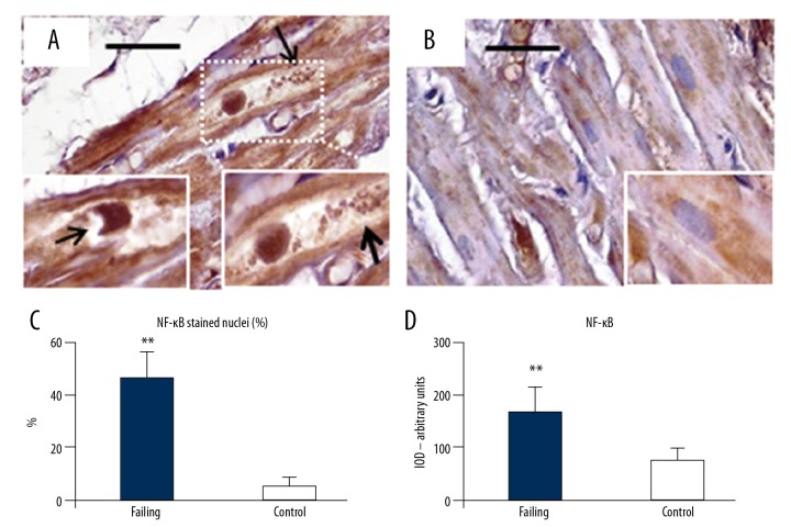
Figure 8.
NF-κB immunostaining. (A) Cardiomyocytes in failing heart had intense NF-κB staining distributed not only inside large cytoplasm areas, but also in a large proportion of nuclei. The cytoplasm appears empty with a lot of debris product (arrows, right insert). The nuclei sometimes showed large “bite-like” signs at pole extremities (arrow in left insert). Conversely (B, and insert), NF-κB was faintly expressed inside the small cytoplasmic areas of control heart samples. The quantitative data of stained nuclei are shows in (C). The optical density for NF-κB staining is resumed in (D). Original magnification 200×; scale bar 30 μm; ** P<0.01. NF-κB – nuclear factor kappa-light-chain-enhancer of activated B cells.

