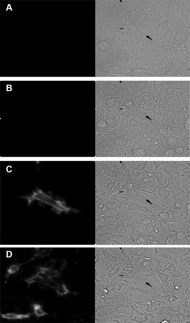
Fig. 8.

Bioluminescent imaging of the apoptotic response. An Olympus LV200 was used to collect phase-contrast and luminescence associated with PS exposure over a 6-h time course of HeLa treated with 2 µM staurosporine. Time zero (a), 3 h (b), 3.7 h (c) and 6 h (d) images shown
