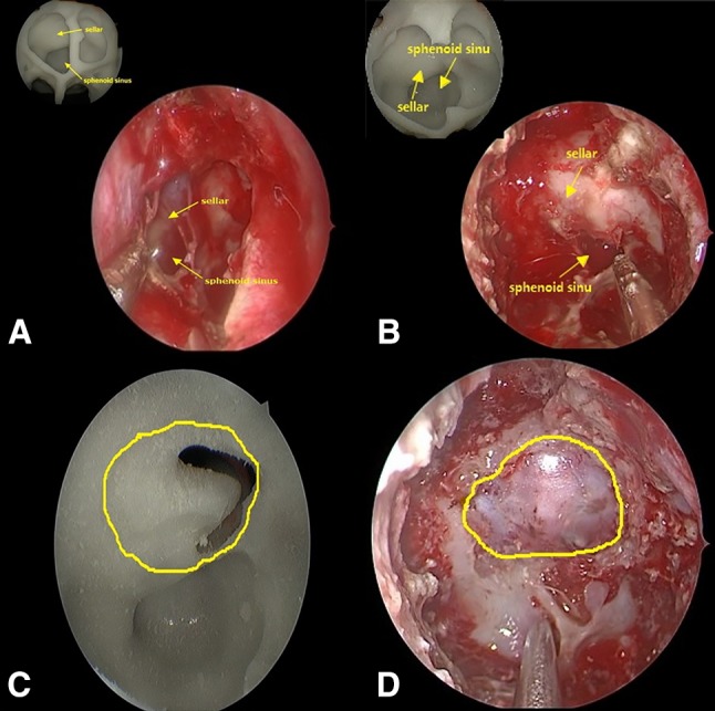
Fig. 3.

a Endoscopy of the anatomical status of the sphenoidal sinus and copmparing with the model. b Exposed sellar floor after removal of the septal bone and copmparing with the model. c Tumor site after removal of the sellar floor bone. d Exposed area of the sellar floor observed in surgery
