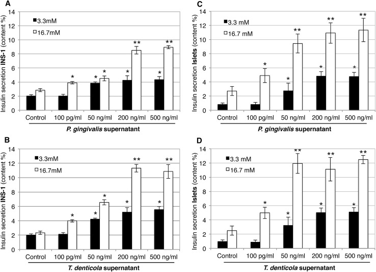
Fig. 3.
Effect of bacterial supernatant treatments of P. gingivalis and T. denticola on insulin secretion in INS-1 β-cells (a, b) and pancreatic islets (c, d) stimulated with 3.3 mM glucose (black bars) and 16.7 mM glucose (white bars). The glucose-stimulated insulin secretion was analyzed by ELISA and insulin secretion was presented as % content, showing similarly increased levels of secreted insulin in INS-1 β-cells and islets infected with P. gingivalis and T. denticola supernatants (50, 200, and 500 ng/ml) in stimulations with both 3.3 and 16.7 mM glucose. x-axis: bacterial species and concentration of bacterial supernatants, y-axis: % content in insulin secretion in INS-1 β-cells and islets. Data are mean ± SD of at least three independent experiments; *p < 0.05, **p < 0.01

