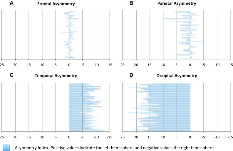
Fig. 5.

Lobar asymmetry retrieved from subtraction of the right side from the left and relating the result to the whole volume of the lobe. The negative values represent the right hemisphere and the positive values the left hemisphere. a Frontal lobe (result non-significant); b parietal lobe (p < 0.003); c temporal lobe (p < 0.0001); d occipital lobe (p < 0.0001)
