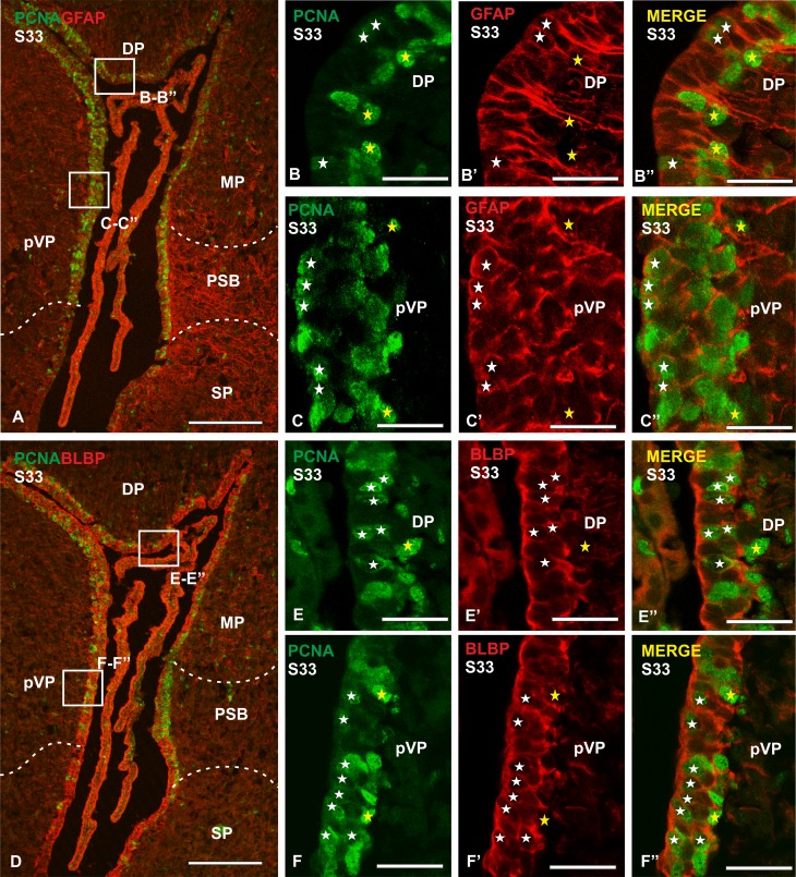
Fig. 8.
Photomicrographs of different sections of the telencephalon of S. canicula in S33 embryos showing double immunofluorescence for PCNA and GFAP or BLBP. a Panoramic view of the telencephalic ventricle showing double immunofluorescence for PCNA and GFAP. b–c′′ Details of the ventricular zone of dorsal pallium and presumptive ventral pallium showing numerous double-labelled cells (white stars) and some BLBP− PCNA+ cells (yellow stars). d Panoramic view of the telencephalic ventricle showing double immunofluorescence for PCNA and BLBP. e–f′′ Details of the ventricular zone of dorsal pallium and presumptive ventral pallium showing numerous double-labelled cells (white stars) and some BLBP− PCNA+ cells (yellow stars). In higher magnification details the ventricle is on the left side of the photomicrographs. Scale bars 200 µm (a, d), 25 µm (b–b′′, c–c′′, e–e′′, f–f′′)

