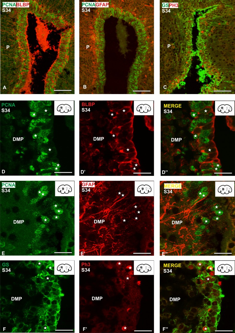
Fig. 9.
Transverse sections of the pallium of a S34 of development (prehatching) at different magnifications showing double immunofluorescence for the three glial markers BLBP, GFAP, GS and the proliferating markers PCNA, PH3. a Panoramic view of the telencephalic ventricle showing double immunofluorescence for BLBP and PCNA. b Panoramic view of the telencephalic ventricle showing double immunofluorescence for GFAP and PCNA. c Panoramic view of the telencephalic ventricle showing double immunofluorescence for GS and PH3. d–d′′ Details of the ventricular zone of a S34 of development showing double-immunolabeled cells for BLBP and PCNA (white stars). e–e′′ Details of the ventricular zone of a S34 of development showing double-immunolabeled cells for GFAP and PCNA (white stars). f–f′′ Details of the ventricular zone of a S34 of development showing double-immunolabeled cells for GS and PH3 (white stars). In higher magnification details the ventricle is on the right side of the photomicrographs. Scale bars: 100 µm (a–c) 25 µm (d–f′′)

