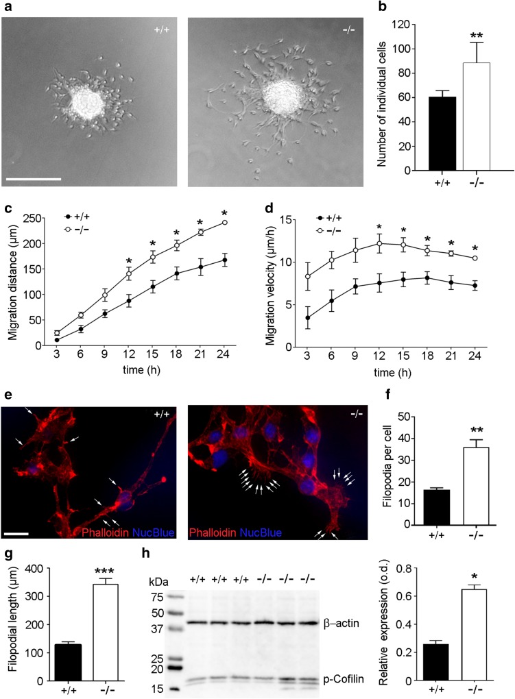
Fig. 4.
Bmal1 deficiency affects migration of NPCs, filopodia morphology and phospho-cofilin expression in vitro. a Representative microphotographs of neurospheres and out-migrating NPCs derived from Bmal1+/+ mice (+/+) and Bmal1−/− mice (−/−). Scale bar = 300 µm. b Quantification of detached NPCs 24 h after seeding. The number of detached NPCs was significantly higher in Bmal1−/− as compared to Bmal1+/+, *P < 0.05, n = 6 mice per genotype. c Migration distance and consequently (d) migration velocity were significantly different between Bmal1+/+ and Bmal1−/− during the first 24 h after seeding *P < 0.05, n = 4 mice per genotype. e Representative photomicrographs of neuroblasts 24 h after seeding with F-actin staining (Phalloidin) and nuclei staining (NucBlue), scale bar 20 µm. Arrows indicate filopodia. f Quantification of filopodia number per cell, **P < 0.01, n = 6 mice per genotype (g) Quantification of filopodial length, **P < 0.01, n = 6 mice per genotype h representative immunoblots and quantification of phospho (p)-cofilin in NPCs from Bmal1+/+ mice and Bmal1−/− mice. *P < 0.05, n = 4 mice per genotype

