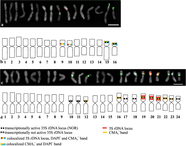
Fig. 4.
Number and localization of 35S rDNA sites (green fluorescence) and 5S rDNA sites (red fluorescence) and idiograms of A. nigrum (a, b) and the triploid A. sphaerocephalon (c, d) with the localization of 35S rDNA, 5S rDNA, NORs, positive CMA3 bands, and positive DAPI bands indicated. All of the chromosomes from the karyotype are presented in the idiograms. Bar 10 μm

