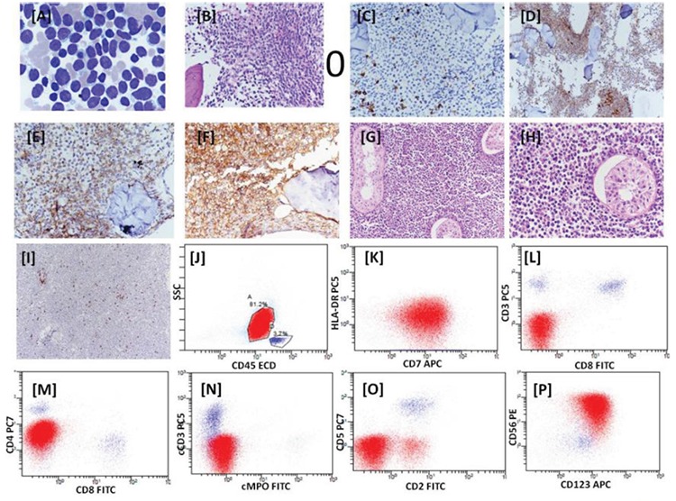
Figure 1.

Bone marrow aspiration showing blast-like cells, Giemsa stain, 100x (A). Bone marrow biopsy showing replacement of marrow by monomorphic cells, hematoxylin and eosin stain, 40x (B). Immunohistochemistry on bone marrow biopsy with CD3 (C), CD7 (D), CD4 (E), and CD43 (F). Histology of testicular mass, hematoxylin and eosin stain, 20x (G), and hematoxylin and eosin stain, 40x (H). Immunohistochemistry of testicular mass with CD3 (I). Dot plots with CD45 vs. SSC showing blast population highlighted in red and lymphocytes in blue (J-P). These dot plots demonstrate the expression of CD7dim, CD4dim, CD56, and CD123. The blasts are negative for CD19, CD10, cCD3, cMPO, CD3, CD8, CD5, CD2, and HLA-DR. 128x96 mm (72x72 DPI).
