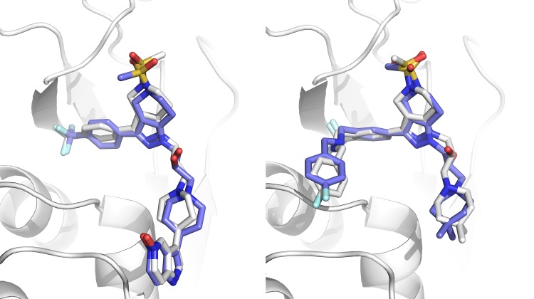
Fig. 2.
Superpositions of HADDOCK models on reference structures. Left: model 5 from target 1 (1.1 Å). Right: model 1 from target 8 (1.5 Å). The reference protein structure is shown in cartoon representation in white. The compounds are shown in stick representation in white and blue for the reference and model molecules respectively. Figure created with PyMOL [29]

