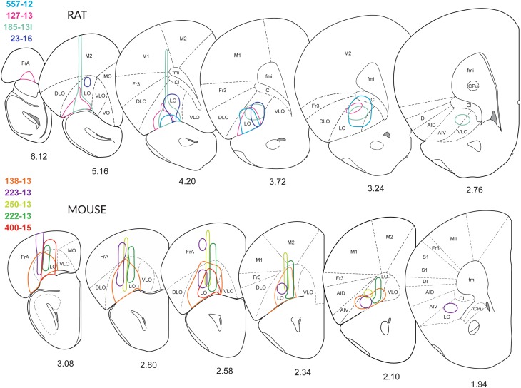
Fig. 2.
Extent of the injections. Schematic drawings of coronal sections of the right hemisphere depicting the extent of the injection sites through the brains of four rats (557-12, 127-13, 158-13 and 23-16) and five mice (138-13, 222-13, 223-13, 250-13 and 400-15), reproduced from (Franklin and Paxinos 2008). The medial boundary of the LO-cortex has been slightly modified, and the VLO-cortex has been introduced according to (Dong 2008; Krettek and Price 1977). In most cases, an adeno-associated virus tracer was injected stereotactically. In cases 250-13 and 400-15, BDA was applied. Although the tracers were injected into the LO-cortex, also dorsal (Fr, Cl and M2) or adjacent regions (DLO and AIV) were sometimes co-labelled. In rats, the injections 557-12 and 127-13 were located mainly in the LO-cortex. In the murine OFC, the injections in the LO-cortex transgressed the border to surrounding areas. In both rats and mice, the tracer injections revealed clearly visible terminal fields in the parvafox-, the Su3- and the PV2 nuclei. The drawings were prepared based on the intrinsic fluorescence of the tracers, in the absence of amplification with antibodies. The drawings are not to scale

