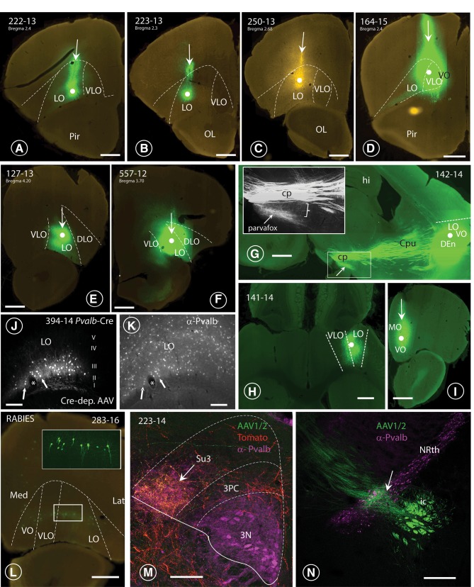
Fig. 3.
Injection sites in the OFC and terminal fields in lateral hypothalamus and PAG. A–F Nanozoomer scans of coronal sections through six specimens (4 mice: A–D; 2 rats: E, F), revealing the course of the Hamilton-syringe needle (vertical arrows) and the site of deposition of the tracer at its tip (white dot). In three cases (A 222-13; E 127-13; F 164-15; G 557-12), the six layers of the OFC-cortex were imbibed with the tracer, whereas in cases B (223-13) and c (250-13), only the deeper layers (V and VI) of the LO-cortex were labelled. In each of the depicted cases, axonal terminals were revealed in the parvafox nucleus, as well as in the Su3- and the PV2 nuclei. G: Para-sagittal section through the brain of a mouse in which the tracer had been injected into the LO-cortex [with spreading to the VLO- and the VO- cortices (142-14)]. The bundles of axons passing through the CPu and converging on the cerebral peduncle (cp) of the posterior hypothalamus are clearly visible (see also Fig. 4a). The terminal field to the parvafox nucleus is indicated with an arrow. The inset shows the almost vertical projection of thin collateral axons (bracket), deriving from the cp and generating the rich terminal field of the parvafox nucleus. H Horizontal section through a mouse brain (rostral side up), injected into the LO-with spreading to the VLO-cortex (141-14). I Coronal section through a mouse brain showing an injection limited to the MO-cortex (129-15), with no projections to parvafox, Su3 and PV2. J, K Sections through the LO-cortex in a Pvalb-Cre mouse (394-14) which had been injected with a Cre-dependent AAV-virus (J). The section was then incubated with an antibody against Parv (K). The cells that took up the tracer are Parv-immunoreactive (arrows). *Blood vessel. L Transsynaptic retrograde visualization of neurons in the prefrontal cortex after Cre-dependent rabies injection in the parvafox nucleus of a Pvalb-Cre/Foxb1-Cre mouse (183-16). Positive neurons are mainly detected in layers V–VI of the LO/VLO cortex. The inset shows an image stack taken with the confocal microscope in the area of the VLO-LO-cortex (frame): the perikarya and the apical dendrites of the pyramidal cells are well visible. Lat lateral, Med medial. M Overlapping terminal fields in the ventrolateral portion of the Su3-region of the PAG (arrow). The green (tracer injected in the LO-cortex) and the red (tracer injected in the parvafox of a Pvalb-Cre/Foxb1-Cre mouse) terminals intermingle and generate the orange tonality (arrow) in Su3. Pvalb-immunoreactivity (magenta) highlights the oculomotor nucleus (3N). 3PC: parvicellular part of the oculomotor nucleus. N Presence of terminals coming from the LO/VLO cortex in the reticular thalamic nucleus (NRth). Scale bars A–D, E, H, I, L, N: 0.5 mm; J, K 0.05 mm; F, G 1 mm; M 0.1 mm

