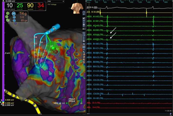
Figure 1.

Top panel shows geometry and voltage map of left atrium in posterior anterior view. The HD Grid multipolar mapping catheter and the ablation catheter are lying on top on the posterior antrum of the left upper pulmonary vein (LUPV). Bottom panel shows the electrogram (EGM) recording via Ensite Velocity 3D mapping system. Solid arrows show local EGM from the LUPV recorded by HD Grid catheter. However, these were not picked up on the ablation bipole
