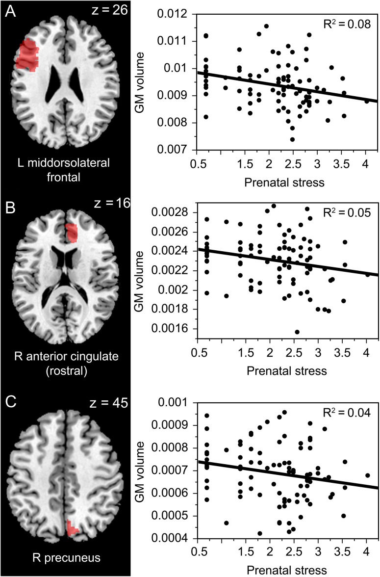
Figure 2.
Prenatal stress and its effect on GM volume (corrected for the total brain volume) in regions identified by Jensen et al. (2015)’s meta-analysis as hypometabolic in depressed patients vs. healthy controls—left mid-dorsolateral frontal cortex ( A; beta = −0.29, P = 0.005, 95% CI [−682.78, −128.27], R2 = 0.08), right anterior cingulate rostral (B; beta = −0.21, P = 0.04, 95% CI [−177.29, −6.01], R2 = 0.05) and right precuneus ( C; beta = −0.20, P = 0.05, 95% CI [−84.69, −0.004], R2 = 0.04) in young adulthood (Prenatal stress refers to the log-transformed mean score on the Stressful life events questionnaire).

