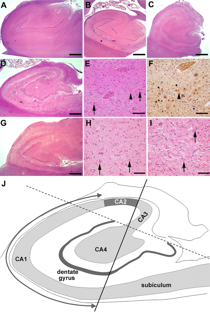
FIGURE 1:
Photomicrographs showing hippocampal sclerosis of Aging (HS-Aging) and frequent comorbidities that mimic hippocampal sclerosis. An 88-year-old male with a macroscopically normal hippocampus and Alzheimer disease neuropathologic change (ADNC) but without HS-Aging (A). A 68-year-old male with frontotemporal lobar degeneration and transactive response DNA-binding protein of 43 kDa (FTLD-TDP) + HS-Aging (B). There is marked atrophy of the CA1 (*). An 89-year-old female with intermediate or high ADNC + HS-Aging (C). There is atrophy, pallor and severe neuronal cell loss and gliosis in CA1. A 95-year-old female with high ADNC and severe neuronal loss mimicking HS-Aging (D-F). There is atrophy and pallor in both the CA1 and the subiculum (D); at higher magnification, several ‘ghost’ tangles are visible (E, F, arrows) and a reactive astrocyte is labeled (arrowhead). A 73-year-old female with FTLD-TDP mimicking HS-Aging (G to I). There is narrowing and pallor not only of CA1 and the subiculum but also of CA2 and CA3 (G). There is severe neuron loss and frequent reactive astrocytes (arrows) in CA1 (H) as well as in the PHG (I). (A - E and G - I) hematoxylin and eosin stain; (F), modified Bielschowsky silver impregnation. Scale bar = 500 μm (A-C, D and G), 100 μm (E, F, H and I). (J) Diagram of the hippocampus used to define hippocampal subfields for neuron number estimation. CA1 = Cornu Ammonis 1 field.

