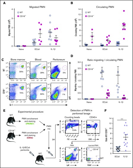
Figure 3.
The dependence of neutrophils on β2 integrins for migration to peritoneum varies with stimulus. (A-B) WT and CD18−/− mice were injected intraperitoneally with E coli (107 CFU per mouse) or IL-1β (5 ng per mouse). Three hours later, PMNs in peritoneum lavage (A) and blood (B) were counted by FACS using counting beads (left histograms). (C) Percentage of Gr-1/Ly6C-high PMNs among CD45+ cells in WT and CD18−/− bone marrow (left), blood (middle), and peritoneum lavage (right) was evaluated by FACS. Representative of at least 5 mice. (D) Ratio of migrated-to-circulating PMNs, allowing an estimation of the relative PMN migration in WT and CD18−/− mice; n = 3-8 mice per group. (E) PMNs were enriched from WT and CD18−/− marrow, stained with PKH67 (green) or CVm (far-red), and mixed at a 1:1 ratio (colors for WT and CD18−/− cells varied across experiments). Cells were injected IV into mice treated with E coli or IL-1β intraperitoneally 1 hour previously. Three hours after cell injection, the presence of green PMNs (WT) and far-red PMNs (CD18−/−) in peritoneal lavage was assessed by FACS. (F) Calculation of the ratio of migrated WT-to-CD18−/− PMNs allows an estimation of the β2 integrin dependency in these 2 models of peritonitis; n = 13 mice per group. ***P ≤ .001. FSC, forward scatter; SSC, side scatter.

