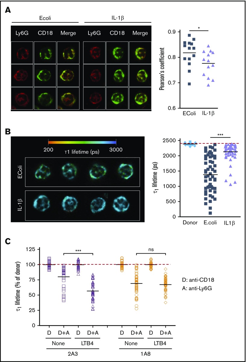
Figure 5.
Ly6G ligation modulates the spatial relationship of Ly6G with β2 integrins. (A) Mice were treated with either E coli or IL-1β intraperitoneally 3 hours later, cells from peritoneal lavage were fixed and stained with Ly6G (AF594, red) and CD18 (FITC, green) monoclonal antibodies (mAbs). (Left) Representative photos from confocal microscope. (Right) Pearson coefficient in PMNs from E coli– or IL-1β–treated mice. (B) Peritoneal PMN from E coli– or IL-1β–treated mice were stained with FITC anti-CD18 (donor) and AF594 anti-Ly6G (acceptor) mAbs. Left, Representative images from each group displaying interacting τ1 lifetime in each pixel on a pseudocolor scale. Right, τ1 lifetime in at least 55 different pixels, representative of 3 independent experiments. Each image in panels A-B represents 12 × 12 μm. (C) PMNs were incubated with 10 μg/mL 1A8 or 2A3 and stimulated or not with 20 nM LTB4. After fixation, cells were stained with anti-CD18 (donor, “D”) without or with anti-Ly6G (acceptor, “D+A”) for FLIM. At least 55 different pixels per conditions were analyzed. Results are expressed as a percentage of τ1 lifetime in donor conditions and are representative of 2 independent experiments. *P ≤ .05; ***P ≤ .001.

