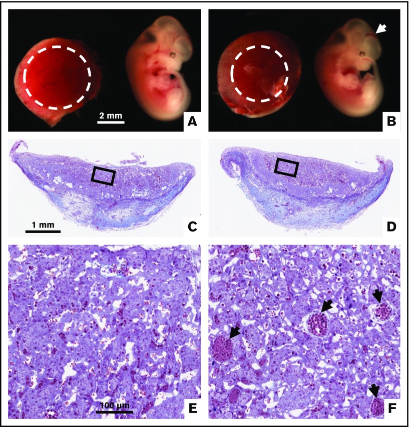
Figure 2.
Intracranial hemorrhage and reduced placental vascularization in global TFPI K1 null embryos generated by TFPI_K1+/−intercrosses. To replicate previously published observations with global deletion of TFPI K1 domain, we examined pregnancies from TFPI_K1+/− intercrosses. Whole-mount images of 11.5 dpc TFPI_K1+/+ (A) and TFPI_K1−/− (B) embryos and placentas are shown. The arrow points to intracranial hemorrhage of the TFPI_K1−/− embryo. Labyrinth regions of placentas are highlighted with dotted circles; the placenta of the TFPI_K1−/− embryo shows reduced labyrinth region. Carstairs’ stained histological sections of both placentas are shown in panels C and D, and boxed regions are enlarged in panels E and F, respectively. Arrows point to large fetal vessels that may not have been optimally branched.

