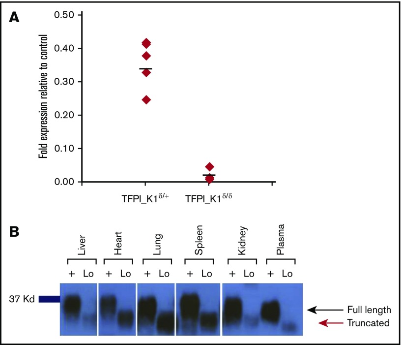
Figure 3.
RNA and protein expression in TFPI_K1δ/δmice. (A) Expression of exon 4 containing RNA in TFPI_K1δ/+ and TFPI_K1δ/δ embryos relative to TFPI_K1Lox/+ litter mates is shown. TFPI_K1δ/δ embryos show 95% to 99% reduction in exon 4 containing TFPI RNA. RNA was isolated from whole embryos. Primers specific for exon 4 were used for real-time qPCR analysis. (B) TFPI_K1δ/δ mice express a truncated protein corresponding to K1-deleted TFPI. FXa-conjugated beads were used to pull down TFPI, and western immunoblotting was performed to evaluate the level of full-length protein in the organs of adult TFPI_K1δ/δ mice (lanes marked “Lo”) and WT C57Bl/6 controls (lanes marked “+”). No full-length TFPI protein could be detected in TFPI_K1δ/δ mice. A truncated protein at reduced level of expression was readily detected.

