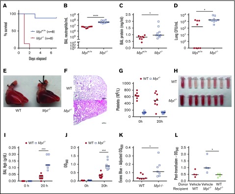
Figure 1.
Thrombocytopenic Mpl−/−mice sustain severe lung injury after IT PA exposure. (A) Mpl−/− and Mpl+/+ mice survival following IT inoculation with PA (P < .0001, log-rank [Mantel-Cox] test). In separate experiments, BAL neutrophil counts/mL (B), BAL protein concentrations (mg/mL) (C), and lung bacterial CFU/mL (D) were measured 20 hours post-PA infection (n = 8 Mpl+/+ mice, n = 9 Mpl−/− mice). Whole-lung images (E) and hematoxylin and eosin–stained lung tissue sections (F; scale bars represent 250 μm) from WT and Mpl−/− mice 20 hours following PA infection. (G) Circulating platelet counts obtained from WT and Mpl−/− mice at 0 and 20 hours post-PA infection. Gross appearance of BAL fluid (H), BAL fluid hemoglobin (Hgb) concentration (g/dL) at 0 and 20 hours (I), BAL fluid OD540 measurements at 0 and 20 hours (J) (n = 5/group at 0 hours, n = 8/group at 20 hours, 1 death in the Mpl−/− group prior to 20 hours). (K) Evans blue dye measurements of lung tissue homogenates at OD620 (adjusted for hemoglobin; n = 7 mice/group). (L) BAL OD540 measurements taken at the 20-hour time point from WT or Mpl−/− mice transfused with either vehicle or WT platelets (109) 1 hour post-PA infection (n = 4 mice/group). For all graphs, each tube or point represents an individual mouse, and the group median is displayed. Statistical comparison by Mann-Whitney U test. *P < .05, ***P < .001, and ****P < .0001.

