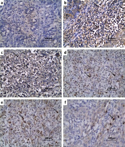
Figure 2.

Renal Sections of Immunohistochemical Staining of NF-κB (p65) (x400) of a) Control Group Showing Minimal Positive Staining for NF-κB (p65); b) Carboplatin group showing strong positive staining for NF-κB (p65) (↑); c) Carboplatin + CMC group showing strong positive staining for NF-κB (p65) (↑);d) Carboplatin + Candesartan group showing moderate positive staining for NF-κB (p65) (↑);e) Carboplatin + CoQ10 group revealing moderate positive staining for NF-κB (p65) (↑); f) Carboplatin + Candesartan + CoQ10 group showing weak positive staining for NF-κB (p65) (↑).
