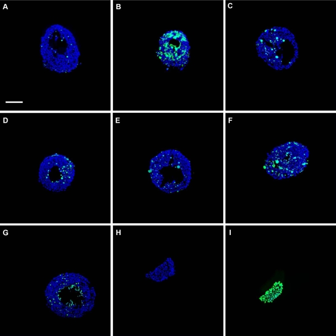
Figure 7.
Representative images of antral follicles subjected to TUNEL. Early antral follicles (n = 6–12 follicles per treatment) were obtained from the ovaries of untreated CD-1 mice, cultured, and processed for TUNEL as described in Materials and Methods section. Representative merged images show DAPI (blue) and TUNEL (green) staining in follicles treated in vitro with vehicle control (DMSO, A), apoptosis positive control (HU, B), DBP 10 μg/ml (C), 100 μg/ml (D), 500 μg/ml (E), 1000 μg/ml (F), and ATBC 0.01 μg/ml (G) for 48 h. Panels H and I show representative images of the assay negative (no enzyme added) and positive (DNAse digestion of tissue) controls, respectively. Images were taken at ×200 magnification. Scale bar represents 100 μm.

