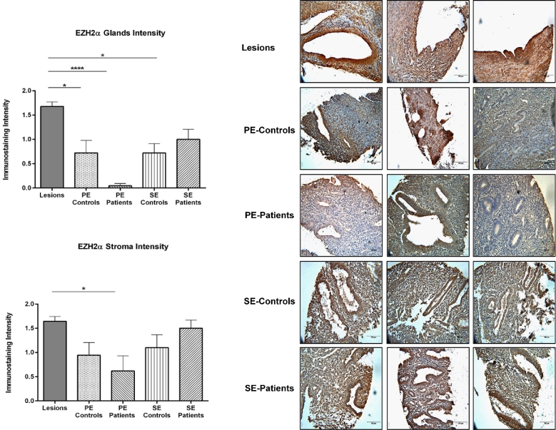
Figure 2.
Immunostaining intensity for EZH2α in endometriotic lesions and endometrium from patients and controls. Immunostaining of EZH2α in glands and stroma was analyzed by three independent scorers using an intensity scale (2 = strong, 1 = weak, and 0 = no staining). Immunostaining was reported as mean intensity ± SEM. Significant higher intensity of EZH2α was observed in the glands and stroma of endometriotic lesions compared to proliferative endometrium (from patients and controls) and secretory endometrium (from patients) (P < 0.05). Representative pictures of EZH2α nuclear immunostaining in lesions and endometria from patients with endometriosis (PE patients) are shown to demonstrate specificity of the antibody and differential expression.

