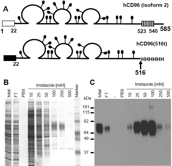
Fig 1. Production of recombinant human CD96.
(A) Schematic representation of human CD96 protein, the 585 amino acid long isoform 2 is represented with residues numbered from methionine 1. The open box represents the signal peptide and the hatched box represents the transmembrane domain. The putative N-linked carbohydrates are shown as black lollipops. In the baculovirus construct, the mellitin signal peptide (black box) replaced the natural signal peptide (amino acids 1 to 21) from CD96. Human CD96 was truncated after lysine 516, and six histidine residues were added at the C terminus of hCD96(516t). (B) Silver stain and (C) western blot analysis of baculovirus-produced hCD96t after nickel chromatography purification and SDS-PAGE under denaturing and reducing conditions. Concentrations of imidazole used to elute proteins from the Ni-NTA-agarose column are indicated. FT = flow through. PBS indicates extensive PBS wash of the column before elution. Sizes of the molecular weight marker are indicated in kilodaltons. Anti-tetraHis Mab (QIAgen) was used to detect tagged hCD96t by western blot (C).

