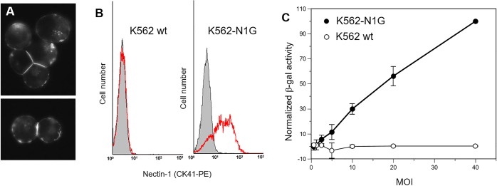
Fig 4. Expression of GFP-nectin-1 in human K562 cells.
(A) K562 cells transiently transfected with plasmid pCK495 express nectin-1 with GFP tagged at the N-terminus. GFP-nectin-1 is expressed at the cell surface and accumulates at cell-cell contacts. Images of GFP fluorescence, magnification 40x. (B) Flow cytometry analysis of K562 cells stably transfected to express GFP-nectin-1. Cells were stained with PE-tagged anti-nectin-1 antibody CK41. Left histogram: K562 cells stained with CK41-PE (red line) are compared to control (gray shade). Right panel: cells from clone K562-N1G #11 expressing GFP-nectin-1 were stained with CK41-PE (red lines vs control shaded in gray). (C) Wild type K562 cells and K562-N1G cells were exposed to lacZ reporter virus HSV-1 KOStk12 at the indicated MOI. Activity of the virus-encoded beta-galactosidase was recorded as the change of OD570nm over time to reflect virus entry. Values from at least two independent experiments were normalized to the highest value (K562-N1G cells at MOI = 40) set at 100% and averaged. Error bars indicate standard deviations across independent experiments.

