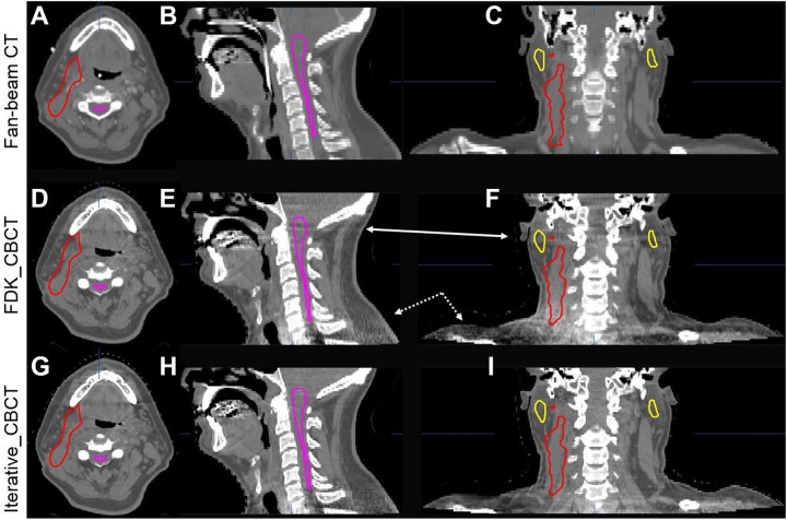
Figure 5.
Comparison of CT images (upper row, A-C) and CBCT images reconstructed by FDK_CBCT (middle row, D-F), and iterative_CBCT algorithm with Medium noise reduction factor (lower row, G-I) and for a head/neck patient. HU window: [−400, 600 HU]. Artifacts (arrows on the coronal and sagittal views) are mitigated in the iterative_CBCT reconstructions. Contours of spinal cord, left and right parotid glands, and target are displayed. CBCT indicates cone-beam computed tomography; CT, computed tomography; FDK, Feldkamp-Davis-Kress.

