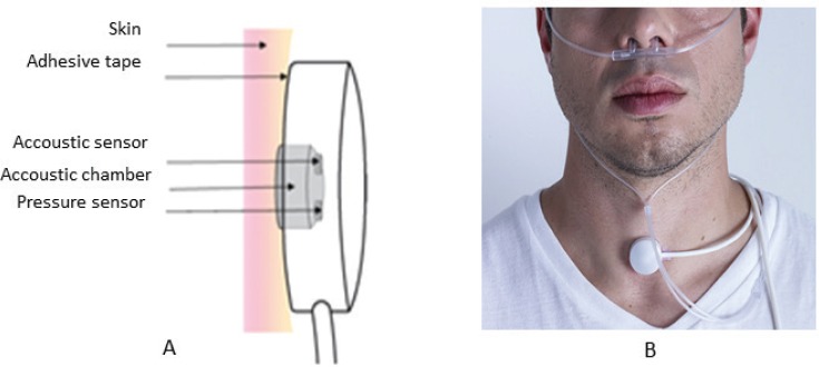
Figure 1. Presentation of a tracheal sound transducer.
(A) Diagram of the PneaVoX sensor that uses both an acoustic sensor and a pressure sensor. The sensors are inserted in a protective plastic housing to ensure an airtight acoustic chamber between the skin and the transducer. (B) Placement of tracheal sound sensor right above the sternal notch using a double-faced tape. If necessary, an adhesive bandage could be used over the sensor to hold it in place.

